Home
/ Diagram Of Plant Cell As Seen Under Electron Microscope / Cell Structures As Seen Under The Light Microscope / (ii) presence of large central vacuole in plant cell.
Diagram Of Plant Cell As Seen Under Electron Microscope / Cell Structures As Seen Under The Light Microscope / (ii) presence of large central vacuole in plant cell.
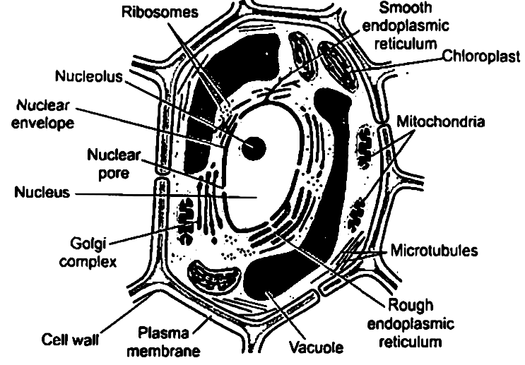
Diagram Of Plant Cell As Seen Under Electron Microscope / Cell Structures As Seen Under The Light Microscope / (ii) presence of large central vacuole in plant cell.. A) name the parts labeled a and b. As you can see from the diagram below plant cells contain a nucleus just as animal cells do. See how a generalized structure of an animal cell and plant cell look with labeled diagrams. Typical animal cell pinocytotic vesicle lysosome golgi vesicles golgi vesicles rough er (endoplasmic reticulum) smooth er (no ribosomes) cell (plasma) membrane mitochondrion golgi apparatus nucleolus nucleus centrioles (2) each composed of 9 microtubule triplets microtubules cytoplasm ribosome Image:plant cell seen under light microscope the cell as seen under the electron microscope.
The diagram is very clear, and labeled; The figure below is a fine structure of a generalized animal cell. (ii) presence of large central vacuole in plant cell. Given alongside are diagrams of plant cells as seen under the microscope after having been placed in two different solutions: Describe how turgor pressure builds up.
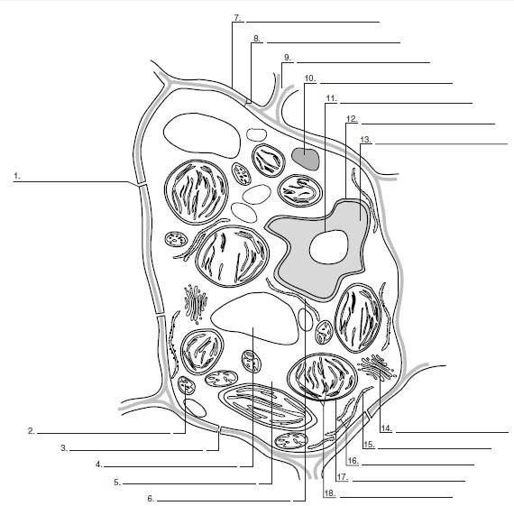
Label The Structures Of A Plant Cell As Seen In A Transmis Chegg Com from media.cheggcdn.com It uses a beam of electrons to illuminate the specimen instead of light as in the case of light microscope. The cell membrane, also known as plasma membrane or plasmalemma consists of three layers when viewed under the electron microscope. Plant cell diagram under electron microscope. Image:plant cell seen under light microscope the cell as seen under the electron microscope. Draw a labelled diagram of the internal structure of a plant cell as seen with an electron microscope. Calculate the actual length of structure c. Here's a diagram of a plant cell: In the cell, there are organelles which are suspended within an aqeuos medium and contained.
See how a generalized structure of an animal cell and plant cell look with labeled diagrams.
(a) based on the diagram, state whether it represents an animal cell or plant cell (b) give two reasons for your answer in (a) above (c) why is the palisade layer a tissue? As seen under a light microscope. The electron microscope is more powerful than the light microscope. In truth, there are still features of plant and anim. A plant cell as seen under electron microscope. (ii) from the solutions given in brackets (water, strong sugar solution, 1% salt solution) name the solution into which : Plant cell structure (jimmy robinson) animal and plant cells have certain. Calculate the actual length of structure c. Though we cannot see everything through the light microscope, some important organelles are visible and we can begin to see some structural differences. Plant cell under the microscope. A plant cell as seen under electron microscope. Describe and compare the structure of a plant cell with an animal cell, as seen under a light microscope, limited to cell wall, nucleus, cytoplasm, chloroplasts, vacuoles and location of the cell membrane. Image:plant cell seen under light microscope the cell as seen under the electron microscope.
The cell membrane, also known as plasma membrane or plasmalemma consists of three layers when viewed under the electron microscope. This will also help you to draw the structure and diagram of plant cell. Endoplasmic reticulum rough and smooth british society for. The diagram below shows the general structure of an animal cell as seen under an electron microscope. Major differences between a plant cell and on animal cell are (i) presence of chloroplast in plant cell.
Biology Questions And Answers Form 1 Biology Form One Notes Ugfacts Net Ke from ugfacts.net Major differences between a plant cell and on animal cell are (i) presence of chloroplast in plant cell. The figure below is a fine structure of a generalized animal cell. See how a generalized structure of an animal cell and plant cell look with labeled diagrams. (i) what is the technical term for the condition of: (iii) presence of cell wall. Bookfanatic89 diagram of plant cell under electron microscope. Draw a labelled diagram of the internal structure of a plant cell as seen with an electron microscope. The cell wall is distinctly visible around each cell.
Cell structure teaching resources the science teacher, organelles biology for majors i, 11 different types of cells in the human body, class test, chronic inflammation under the microscope learn share.
But at the same time it is interpretive. The diagram below represents a cell as seen under an electron microscope. The diagram below shows the general structure of an animal cell as seen under an electron microscope. The structures within the cell are referred to as organelles. (iii) presence of cell wall. Robert hooke in 1665 first discovered plant cell. Click (or tap) the diagram for a simple labelled version. When viewing onion cells under a microscope a few drops of a certain solution are added to stain the cells and show these cells more clearly. (ii) presence of large central vacuole in plant cell. Plant cell diagram under electron microscope. A) name the parts labeled a and b. See how a generalized structure of an animal cell and plant cell look with labeled diagrams. The cell membrane, also known as plasma membrane or plasmalemma consists of three layers when viewed under the electron microscope.
As you can see from the diagram below plant cells contain a nucleus just as animal cells do. As seen under a light microscope. The diagram is very clear, and labeled; The diagram below shows the general structure of an animal cell as seen under an electron microscope. Typical animal cell pinocytotic vesicle lysosome golgi vesicles golgi vesicles rough er (endoplasmic reticulum) smooth er (no ribosomes) cell (plasma) membrane mitochondrion golgi apparatus nucleolus nucleus centrioles (2) each composed of 9 microtubule triplets microtubules cytoplasm ribosome
Long Answer Question Illustrate Only A Plant Cell As Seen Under Electron Microscope How Is It Different From Animal Cell Snapsolve from wb-qb-sg-oss.bytededu.com The structures within the cell are referred to as organelles. When viewing onion cells under a microscope a few drops of a certain solution are added to stain the cells and show these cells more clearly. The figure below is a fine structure of a generalized animal cell as seen under an electron microscope. Describe and compare the structure of a plant cell with an animal cell, as seen under a light microscope, limited to cell wall, nucleus, cytoplasm, chloroplasts, vacuoles and location of the cell membrane. A) name the parts labeled a and b. Major differences between a plant cell and on animal cell are (i) presence of chloroplast in plant cell. The cell wall is distinctly visible around each cell. This will also help you to draw the structure and diagram of plant cell.
Typical animal cell pinocytotic vesicle lysosome golgi vesicles golgi vesicles rough er (endoplasmic reticulum) smooth er (no ribosomes) cell (plasma) membrane mitochondrion golgi apparatus nucleolus nucleus centrioles (2) each composed of 9 microtubule triplets microtubules cytoplasm ribosome
But at the same time it is interpretive. The structures within the cell are referred to as organelles. Some of the cell organelles that can be observed under the light microscope include the cell wall, cell membrane, cytoplasm, nucleus, vacuole and chloroplasts. B) how the is structure labeled b adapted to its function? (ii) from the solutions given in brackets (water, strong sugar solution, 1% salt solution) name the solution into which : Electron microscope can magnify an object up to 500, 000 times. A cell is a very tiny structure which exists in living bodies. The figure below is a fine structure of a generalized animal cell as seen under an electron microscope. The electron microscope is more powerful than the light microscope. Three layers together measure approximately 75 å in thickness. In plant cells, the cell membrane lies inside the cell wall. The cell organelles are seen as tiny dots throughout the cytoplasm, whereas the nucleus is seen as a thick drop. A plant cell as seen under electron microscope.
Post a Comment
for "Diagram Of Plant Cell As Seen Under Electron Microscope / Cell Structures As Seen Under The Light Microscope / (ii) presence of large central vacuole in plant cell."


Post a Comment for "Diagram Of Plant Cell As Seen Under Electron Microscope / Cell Structures As Seen Under The Light Microscope / (ii) presence of large central vacuole in plant cell."